


Raynaud’s Phenomenon causes blood vessels to spasm, temporarily restricting blood flow to extremities such as fingers and toes, which can leave them visibly pale and cold. This condition is estimated to affect 20% of the global adult population, including up to 10 million in the UK.[1] Similarly, Hand Arm Vibration Syndrome (HAVS), sometimes known as vibration white finger, occurs when nerves, blood vessels, and joints are damaged due to long-term exposure to vibrations. This results in restricted circulation and temperature drops in the hands and feet. It’s thought that nearly two million people in the UK are at risk of HAVS.[2] Thankfully, thermography is showing promise as a non-invasive screening technique.[3] This is not the first time thermal cameras have been used as a preliminary assessment tool. Previously, they've provided a non-contact method of detecting elevated skin temperature, a non-invasive way to pinpoint the source of cattle lameness, and a non-destructive technique for assessing tree health. These applications highlight the broader potential of thermography as a rapid, non-contact method of identifying physiological change.
Quick Links
- What is the Difference Between Raynaud’s Phenomenon and HAVS?
- How Does Thermal Imaging Show the Impacts of HAVS and Raynaud’s Phenomenon?
- Which Other Conditions Can Thermal Cameras Detect?
- Further Information
What is the Difference Between Raynaud’s Phenomenon and HAVS?
Raynaud’s Phenomenon and Hand Arm Vibration Syndrome (HAVS) share many of the same symptoms, including pain, numbness, and pins and needles in the fingers and toes. During episodes of HAVS or Raynaud’s, extremities may turn white or blue before becoming red and painful as circulation returns.
Often triggered by cold temperatures, stress or anxiety, there are two variations of Raynaud’s Phenomenon:
- Primary: this version of Raynaud’s occurs spontaneously. Primary Raynaud’s could be inherited and is usually relatively mild; it may even disappear in later life, though this is not a guarantee.
- Secondary: typically, much more serious, and much less common, secondary Raynaud’s tends to be associated with underlying health conditions or external factors; HAVS is one such condition that could trigger secondary Raynaud’s Phenomenon.[4]
Unlike primary Raynaud’s Phenomenon, HAVS is irreversible. It results from prolonged and consistent use of vibrating tools, leading to nerve, blood vessel, and joint damage in the hands and arms. The condition usually affects professionals in sectors where high-vibration tools, such as jackhammers, grinders, breakers, jigsaws, lawnmowers, and handheld power tools, are commonly used, for example, construction, mining, engineering, shipbuilding, and forestry.
Restricted blood flow due to damaged capillaries means HAVS presents similarly to Raynaud’s Phenomenon. Unfortunately, joint and nerve injury means HAVS sufferers may also experience the following additional symptoms:
- Loss of touch sensitivity
- Lack of dexterity and/or grip strength[5]
Reducing the Risk of HAVS
Sadly, once contracted, HAVS is irreversible. Worse still, the Health and Safety Executive (HSE) reported 215 new cases of HAVS in 2023.[6] However, according to the Health and Safety at Work Act 1974, employers are required to assess and control risks from vibration exposure. As such, there are several steps that employers and employees can take to reduce the risk of HAVS:
- Use vibration measurement tools to assess risk-levels and degrees of exposure
- Use an alternative method of work that reduces vibration
- Provide hand tools that are vibration-reduced
- Make sure tools are regularly serviced and maintained
- Provide training to prevent bad tool use
- Supply effective PPE such as appropriately rated anti-vibration gloves (please note anti-vibration gloves should not be relied upon as a first or only measure as they provide limited protection)
- Ensure employees have adequately warm clothing if working outdoors or in cold environments
- Encourage employees to report early symptoms[7]
How Does Thermal Imaging Show the Impacts of HAVS and Raynaud’s Phenomenon?
Temporary spasms (Raynaud’s Phenomenon) in blood vessels and damage to capillaries (HAVS) restrict circulation to extremities, such as hands, fingers, feet, and toes. Lack of blood causes the temperature of these body parts to decrease. Thermal cameras detect the intensity of IR energy (heat) emitted across hands and arms, converting it into a visible heat map. As a result, you can see the cooling extremities of Raynaud's and HAVS sufferers.
In fact, in 2016, an IR image depicting the thermal difference between a healthy hand and that of a Raynaud’s sufferer won the Wellcome Trust’s Image Award. The competition sought “to recognise the creators of the most informative, striking and technically excellent images that communicate significant aspects of biomedical science.”[8]

The then chair of the judging panel and head of Wellcome Images, Catherine Draycott, explained that the image, captured by Mathew Clavey, won the prestigious award because:
“[…] it [the thermal image] shows so vividly the difference between normal circulation and the poor circulation of someone with Raynaud’s disease – triggered by cold temperature, stress and anxiety.”[9]
~ Catherine Draycott, Chair of Judging Panel and Head of Wellcome Images
PASS Assesses the Efficacy of Thermal Imaging for Detecting Raynaud’s Phenomenon
Conclusions about the efficacy of thermal imaging as a diagnostic tool for Raynaud’s Phenomenon and HAVS vary between studies (see footnote 3 for details of scientific papers). Nevertheless, infrared thermography is listed as a diagnostic method, alongside other tests, in both NHS and Scleroderma & Raynaud’s UK literature. [10]
In light of this, PASS Ltd decided to conduct our own rudimentary test, using FLIR Edge and FLIR Exx Thermal Cameras. First, to discover whether thermal cameras could illustrate the difference between a healthy hand and that of a Raynaud’s sufferer. Second, to observe the difference in clarity and detail between the two thermal cameras.
Capturing images with both cameras, we compared Katie's hands (our resident Raynaud’s sufferer and Marketing Manager) with Daisy's (our healthy Marketing Apprentice) and Lewis' (our healthy Account Manager) hands.
FLIR Edge Wireless Thermal Camera

In the thermal images, Katie’s fingers appear purple-black, showing that they are significantly colder than the rest of her body, which appears orange-yellow. Her fingers are also notably darker than Daisy’s fingers.

We may suppose that, given Katie suffers from Raynaud’s Phenomenon and Daisy does not, restricted blood flow has caused Katie’s extremities to cool.

This comparison illustrates how thermal imaging can visualise temperature differences associated with restricted circulation.


FLIR Exx Thermal Camera

Like the FLIR Edge, the FLIR Exx Thermal Camera illustrated the difference between healthy hands (Lewis’) and those of someone with Raynaud’s Phenomenon (Katie’s).

The added sensitivity of the FLIR Exx has also enabled it to detect that Katie’s nose is colder than the rest of her body, though not as cold as her hands. A cold nose is another symptom of Raynaud’s Phenomenon.

Moreover, the Exx’s higher IR resolution has rendered the image in greater detail, making it easier to identify features and see the extent of Katie’s restricted bloodflow.


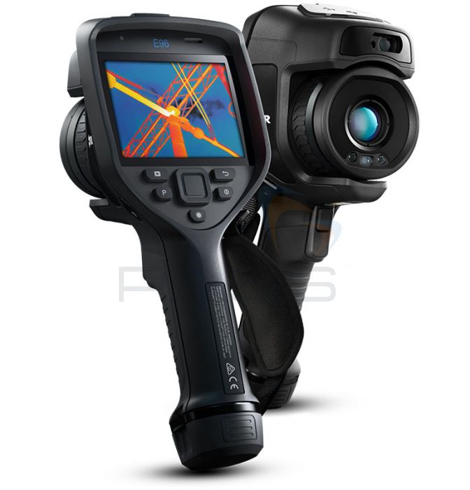
It is important to note that thermal imaging cannot be used in isolation to diagnose Raynaud's Phenomenon or HAVS. However, thermal cameras can visualise the thermal patterns that result from restricted circulation, making thermography a rapid, non-contact method of identifying physiological changes that may warrant further investigation.
Which Other Conditions Can Thermal Cameras Detect?
As demonstrated above, thermal cameras can illustrate the decreased skin temperature associated with Raynaud's Phenomenon and HAVS. They also work in the reverse and are able to detect elevated skin or surface temperatures that are often symptomatic of illnesses in humans and animals. Regardless of whether you are identifying cooling caused by restricted circulation or heat generated by inflammation, thermal imaging makes it easy to identify temperature patterns that reflect underlying physiological change.
The following application stories illustrate how thermal cameras can be used as a preliminary precaution for identifying potential signs of illness. Please note that raised or reduced skin temperatures detected using a thermal camera must be verified using a medical thermometer. Furthermore, it is important to realise that thermal cameras cannot be used in isolation to diagnose conditions, as a change in skin temperature alone is not conclusive proof of illness.
Care Home Resists Coronavirus Using Hikvision Thermal Camera Supplied by PASS Ltd

At the very beginning of the COVID-19 pandemic, there was no vaccine, no real understanding of how to treat cases, and care home deaths were rising. One symptom of coronavirus was an elevated skin temperature. Therefore, to protect their vulnerable, elderly residents, Carlton House care home in New Milton invested in a Hikvision Temperature-screening Thermal Camera, supplied by PASS Ltd. The IR imager was used, not to diagnose COVID-19, but to identify those with a raised skin temperature and, therefore, might be displaying symptoms of the disease. These individuals could then be referred for further tests. Consequently, at the time of writing (16th May 2020), Carlton House had not suffered any coronavirus cases or deaths during lockdown.
This application story illustrates how thermal cameras can be used to detect changes in body temperature that might be indicative of illness, providing a fast method of screening people for additional tests.
Thermal Camera Functions as an Additional Preventive Measure in Care Home During Lockdown
Catch Cattle Lameness with Economical Thermal Cameras

FarmingUK estimates that lameness affects 20% to 25% of UK cattle and costs the dairy industry £53 million a year.[11] Infection or inflammation is usually the root cause of lameness. As both conditions emit heat as a symptom, thermal cameras can be used to pinpoint the location of the lameness and understand its root cause, as livestock manager George Coles explains:
“Straight away you could find that if you had a lame cow, you could see where the hotspots were. [...] The hotspots were often higher up in the leg rather than the foot, so you could treat it with anti-inflammatories, not antibiotics […] Before thermal imaging, my treatment for anything limping was to give them antibiotics and anti-inflammatories and you’ve covered them. Since I’ve been doing this, the only animals that have antibiotics are those that have a case of foul of the foot.”[12]
~ George Coles, Livestock Manager
Identifying the source of lameness earlier means it can be treated effectively using a suitable intervention. This lowers medical costs, reduces animal discomfort, and prevents antibiotic resistance.
Thermal Cameras Save Dairy Farmers Millions in Cattle Lameness
Thermal Cameras Used to Monitor Tree Health
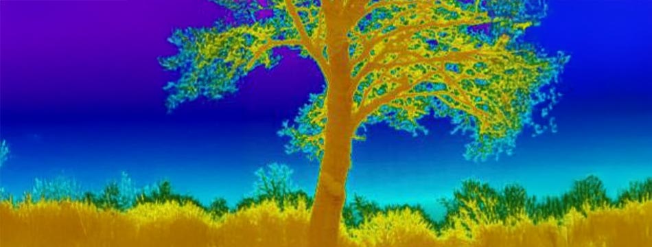
Previously, assessing the health of trees relied upon penetrating the bark. It was time-consuming, provided limited insights, and put the tree at risk of disease. Tree thermography has proven revolutionary. It’s much quicker, offers a holistic view of the tree’s condition, and eliminates pathogen transference between specimens.
The thermal conductivity of wood depends on tissue moisture. As such, cavities, where there is no tissue, or areas that have decayed and are not absorbing as much water, will have reduced thermal conductivity. Consequently, these areas will appear colder on a thermal image compared to healthy tree tissue. This allows conservationists to identify potentially sick trees quickly and easily, enabling early diagnosis and treatment.
Trees Protected from Disease Using Thermal Cameras
Further Information
For additional help or advice regarding our thermal cameras, including models by leading brands such as FLIR and Hikvision, as well as their potential for preliminary screening applications, please contact our Sales team on 01642 931 329 or via our online form.
In the meantime, you can browse our complete collection of thermal cameras here.
Explore Our Range of Thermal Cameras that can Support Human, Animal, and Botanical Health and Welfare Assessments
Footnotes
[1] NHS Inform, Raynaud’s Phenomenon, last accessed 27 February 2026.
[2] Iain Hoey, ‘Bad vibrations: BSIF highlights syndrome affecting UK workforce’, International Fire & Safety Journal, last accessed 27February 2026.
[3] Multiple scientific papers, articles, and organisation resources detail the use of thermography as a diagnostic tool for Raynaud’s Phenomenon or HAVS. Conclusions about the effectiveness of using thermal cameras to identify symptoms of and diagnose primary and secondary Raynaud’s Phenomenon and HAVS in patients vary.
- Ariane L. Herrick, Graham Dinsdale, Andrea Murray, ‘New perspectives in the imaging of Raynaud’s phenomenon’, European Journal of Rheumatology, 7.3 (2020), last accessed 27 February 2026.
- Ronald House, Linn Holness, Ihor Taraschuk, Rosane Nisenbaum, ‘Infrared thermography in the hands and feet of hand-arm vibration syndrome (HAVS) cases and controls’, International Journal of Industrial Ergonomics, 62 (Nov 2017), 70-76, last accessed 27 February 2026.
- Jason Laday, ‘Infrared thermography reliable in assessing children with Raynaud’s phenomenon’, Healio Rheumatology, last accessed 27 February 2026.
- M.J. Lim, S.R. Kwon, K.H. Jung, K. Joo, S.-G. Park, W.Park, ‘AB0739 Infrared thermography of big toe in raynaud’s phenomenon’, Annals of Rheumatic Diseases, 72.3 (June 2013), last accessed 27 February 2026.
- Lotte Lindberg, Bent Kristensen, Ebbe Eldrup, Jane Frølund Thomsen, Lars Thorbjørn Jensen, ‘Infrared Thermography as a Method of Verification in Raynaud’s Phenomenon’, Diagnostics, 11.6 (2021), last accessed 27 February 2026.
- Royal United Hospitals Bath NHS, Thermography and Capillaroscopy, last accessed 27 February 2026.
- Harshwardhan Patil, I.N. Monisha, M.S. Mallikarjunaswamy, P. Praveen, M.S. Sowmya, M. Nidhis Chandra, Mahabaleshwar Mamadapur, ‘Infrared thermography for the diagnosis and monitoring of Raynaud’s phenomenon: current evidence and future directions’, Reumatologia, 63.6 (2025), 416-425, last accessed 27 February 2026.
- Scleroderma & Raynaud’s UK, Raynaud’s Phenomenon, last accessed 27 February 2026.
[4] Information about Raynaud’s Phenomenon was gathered from the following sources:
- NHS, Raynaud’s, last accessed 27 February 2026.
- NHS Inform, Raynaud’s Phenomenon.
- Callum Wallis, ‘What Is HAVS? Causes, Symptoms and Employer Responsibilities’, Pulsar Instruments, last accessed 27 February 2026.
- Scleroderma & Raynaud’s UK, Raynaud’s Phenomenon.
[5] Information about HAVS was gathered from the following sources:
- Iain Hoey, ‘Bad vibrations: BSIF highlights syndrome affecting UK workforce’.
- HSE, Hand-arm vibration, last accessed 27 February 2026.
- Stephen Hussey, ‘What is the Difference Between Raynaud’s Phenomenon and Vibration White Finger?’, The Compensation Experts, last accessed 27 February 2026.
- Callum Wallis, ‘What Is HAVS? Causes, Symptoms and Employer Responsibilities.’
[6] Iain Hoey, ‘Bad vibrations: BSIF highlights syndrome affecting UK workforce’.
[7] Suggestions for reducing the risks of HAVS were gathered from the following sources:
- BSIF, Bad Vibrations – Hand Arm Vibration Syndrome (HAVS), last accessed 27 February 2026.
- Iain Hoey, ‘Bad vibrations: BSIF highlights syndrome affecting UK workforce’.
- HSE, Hand-arm vibration.
[8] Raynaud’s Association, Thermal Image of Raynaud’s Is an Award Winner, last accessed 27 February 2026.
[9] Raynaud’s Association, Thermal Image of Raynaud’s Is an Award Winner.
[10] Royal United Hospitals Bath NHS, Thermography and Capillaroscopy, and Scleroderma & Raynaud’s UK, Raynaud’s Phenomenon.
[11] FarmingUK, Study finds low-cost thermal devices effective in detecting lameness, last accessed 27 February 2026.
[12] Hayley Chapman, 'Thermal imaging cuts antibiotics use on Northants beef farm', Farmers Weekly, last accessed 03 March 2026.